When Bad Things Happen to Great Vessels – Part I
Frank J. Edwards, MD
The aorta, as everyone knows, is a high-pressure, semi-elastic conduit coming off the heart’s left ventricle that arches downwards, dives through the diaphragm and courses through the abdomen into the pelvis, where it bifurcates into the iliac arteries. Major arteries branch off throughout its long course supplying our vital structures with oxygen and nutrients. The wall of the aorta has three layers under the microscope—a strong, fibrous outer layer, a muscular middle layer, and a relatively thin and delicate inner membrane.
When something goes wrong with the aorta, it’s going to be a clinical nightmare. Bullets and blades account for most traumatic injuries, but the aorta can rip when the heart is wrenched and twisted during the first seconds of a high velocity accident or fall. Patients with traumatized aortas usually don’t make it to the hospital. If they do, the challenge is not one of recognizing the problem—
But of fixing it . . . very quickly.
Non-traumatic aortic crises, however, can be surprising difficult to diagnose, and are just as potentially lethal. Such patients may slip through triage looking like back strains, angina, kidney stones, strokes and even constipation.
The nature of non-traumatic aortic catastrophes will vary depending upon location, but fall into two general categories: the thoracic aorta tends to suddenly dissect, while the abdominal aorta will gradually develop aneurysms that enlarge and eventually rupture. Today we’ll look at thoracic aortic dissections, and next month the ruptured AAA (abdominal aortic aneurysm).
 |
| http://www.yalemedicalgroup.org/stw/Page.asp?PageID=STW025691 |
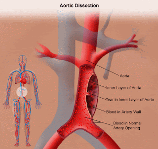
The thoracic aorta is that segment running from the heart to the diaphragm. A dissection occurs when the inner lining develops a sudden, spontaneous tear, which can occur for a number of reasons, including long-standing high blood pressure, congenital connective tissue disorders like Marfan’s Syndrome, and the Ehlers-Danlos Syndrome.
The tear may occur close to the heart (the aortic root) or anywhere further along the vessel as it arches down. Suddenly, all that blood pulsing out of the heart under high pressure has somewhere else to go, and because the toughest outer coating usually holds, it dissects a new channel between the inner and outer layers, and it does so with a vengeance, wrecking havoc along the way.
It can seep toward the heart and block the coronary arteries—giving heart attack symptoms—or it can compromise blood flow to the brain and resemble a stroke. Not uncommonly it will dissect all the way down the length of the aorta into the pelvis and throttle blood flow to one of the iliac arteries, causing pain and numbness in a leg. Furthermore, the outer layer may crack open and allow blood to gush out into the chest cavity. We are talking serious badness anyway you slice the cake.
While a good number of patients with thoracic aortic dissections have severe upper back pain often described as “tearing” in nature, many don’t. They may have only chest pain accompanied by EKG changes resembling a myocardial infarction, or they may have stroke-like neurologic deficits—up to and including coma—or they may have back-pain-plus-leg-numbness, or chest-pain-plus-arm-pain, or back-pain-and-chest pain, or . . . you get the idea. Chest x-ray alone won’t make the diagnosis, and neither will any single test except a contrast enhanced chest CT scan.
This is what happened to the actor John Ritter. He developed nausea and vomiting while on a set and went across the street to a very good medical center in L.A. where an EKG suggested he was having a heart attack. The highly skilled cardiologist on duty ordered a blood thinner and Mr. Ritter died.
Once in a while, thoracic dissections will “stabilize.” The rent will seal, the dissection cease, and the patient may require only good blood pressure control. But treatment usually falls to the thoracic surgeon, and the already high mortality rate rises the longer diagnosis is delayed.
The stop watch has begun ticking before you even lay eyes on the patient.
Emergency medicine providers must keep a high index of suspicion in anyone who complains of upper back pain or chest pain—and especially (here’s an excellent rule of thumb) if the patient has symptoms both above and below the diaphragm. Next month—the equally dreaded “triple A.”
*****************************************************************************
Frank Edwards was born and raised in Western New York. After serving as an Army helicopter pilot in Vietnam, he studied English and Chemistry at UNC Chapel Hill, then received an M.D. from the University of Rochester. Along the way he earned an MFA in Writing at Warren Wilson College. He continues to write, teach and practice emergency medicine. More information can be found at http://www.frankjedwards.com/.


No comments:
Post a Comment